

VitroGel® Hydrogel Matrix
Overview
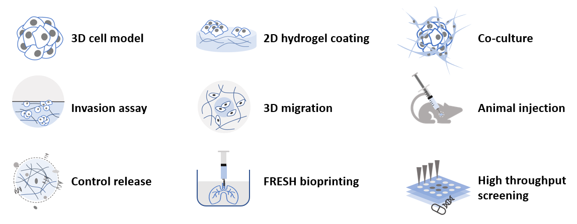
VitroGel® Hydrogel Matrix is a ready-to-use, xeno-free functional hydrogel for 3D cell culture research. VitroGel Hydrogel Matrix is an optimized formulation of multi-functional ligands and concentration to support a wide range of cell types for different applications.
“Just Add Cells” – The hydrogel matrix is ready to mix with cell suspension directly. There is no additional adjustment needed.
VitroGel Hydrogel Matrix closely mimics the natural extracellular matrix (ECM) environment to make cells feel more like at home. The hydrogel is room temperature stable, has a neutral pH, transparent, permeable and compatible with different imaging systems. The solution transforms into a hydrogel matrix by simply mixing with the cell culture medium. Cells cultured in this system can be easily harvested out with our VitroGel Cell Recovery Solution.
This user-friendly functional hydrogel creates an excellent balance of simplicity and versatility.
Specifications
| Formulation | Xeno-free, functional hydrogel |
| Use | 3D and 2D cell culture |
| Operation | Ready-to-use at room temperature |
| Biocompatibility | Biocompatible, safe for animal studies |
| Injection | Injectable hydrogel for in vivostudies and lab automation |
| Cell Harvesting | 20 min cell recovery using VitroGel Cell Recovery Solution |
| pH | Neutral |
| Storage | Store at 2-8°C. Ships at ambient temperature |
| Sizes | 10 mL and 2 mL |
| Usage | 300 uses (10 mL), 60 uses (2mL) |
3D cell culture process in 20 min – “Just add cells”
VitroGel Hydrogel Matrix is ready-to-use. Just mix with your cells. There is no cross-linking agent or the need to adjust the hydrogel concentration.

Handbooks and Resources
Product Documentation
Data and References
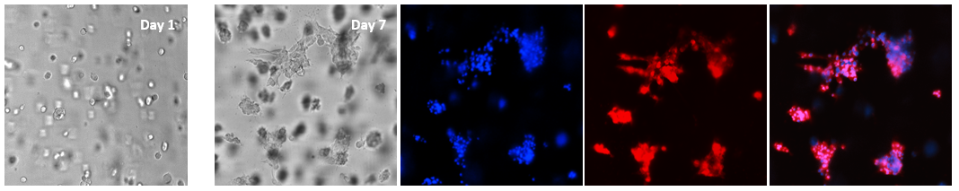
Figure 1. 3D culture of glioblastoma cells (U-87 MG) in VitroGel Hydrogel Matrix
The single-cell suspension was prepared with 50% FBS and mixed with VitroGel Hydrogel Matrix at 4:1 v/v ratio (400 µL VitroGel + 400 µL cell suspension) for the final 10% FBS in the mixture. The cells were cultured in the hydrogel matrix for 7 days and stained with live-cell imaging dyes. The nucleic acid dye (Hoechst 33342, Blue) and cell membrane dye (Red) were added directly in the cover medium and incubated for 30 minutes at 37°C before imaging. Z-stack imaging system with 2D image projection was used for both transmitted light and fluorescent images. The glioblastoma cells grew in the hydrogel matrix and formed 3D colonies. The cell-cell interactions were showed in the extended structure that connected different colonies. The VitroGel Hydrogel Matrix shows great support for 3D glioblastoma cell models with strong cell-cell communications. The hydrogel is extremely easy for image analysis, e.g, by adding the molecular probes directly on the top of the hydrogel.
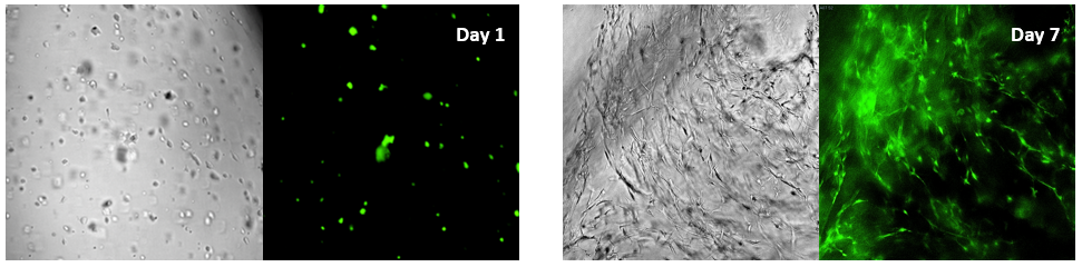 Figure 2. Bone marrow cells 3D cultured in VitroGel Hydrogel Matrix
The fibroblast-like mouse bone marrow stromal cells (OP9-GFP) were 3D cultured in VitroGel Hydrogel Matrix. The single cells were homogenously suspended within the hydrogel matrix and started to form the stretched fibroblast-like structure on day 1. The multiple integrin-binding ligands and cellular functional ligands on VitroGel Hydrogel Matrix can promote a strong cell-matrix interaction, which accelerated the cell proliferation and cell-cell communications during the 3D cell culture. On day 7, a clear 3D cellular networking structure formed within the hydrogel matrix.Z-stack imaging system with 2D image projection were used for both transmitted light and GFP fluorescent images.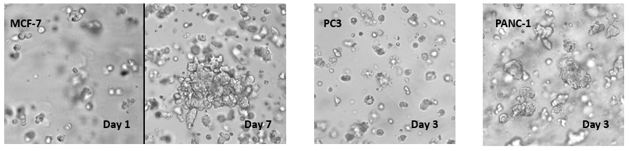
Figure 3. Cancer cells grown in ViroGel Hydrogel Matrix
Various cancer cells 3D cultured in VitroGel Hydrogel Matrix can grow rapidly and form tumor-like structures. The images above show human mammary breast cancer cells (MCF-7), human prostatic cancer cells (PC3) and human pancreatic cancer cells (PANC-1) 3D cultured in VitroGel Hydrogel Matrix. The cells were prepared as single cells suspension and encapsulated within the hydrogel matrix. The grape-like shaped cell colonies in MCF-7 cells starts to form (day 1). The cell colonies continue to grow and formed the tumor-like structure (day 7). A similar phenomenon was also presented in PC3 and PANC-1 cells where the cells grew rapidly into a 3D mini-tumor within the hydrogel matrix.